If sarcoma starts in smooth muscle, it is called leiomyosarcoma.
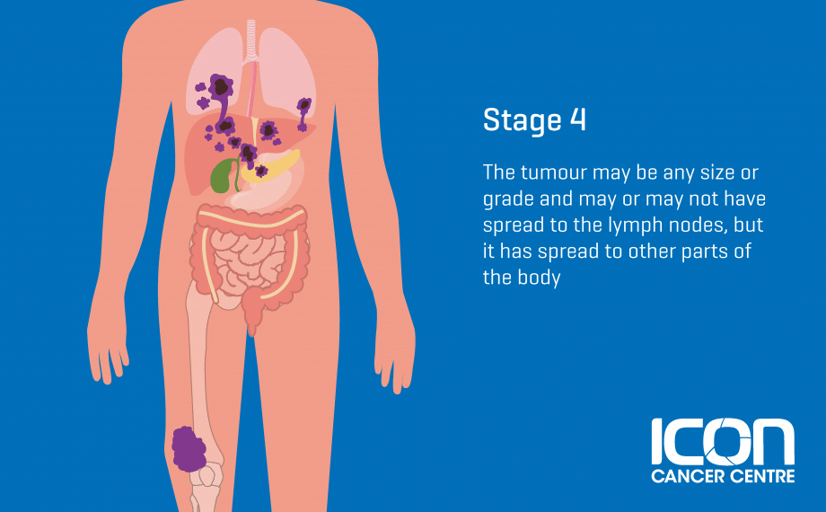
sarcoma abdominal Although there are more than 50 types of sarcoma, they can be grouped into two main kinds: A sarcoma is a type of cancer that starts in tissues like bone or muscle. Soft tissue sarcoma is a rare type of cancer that starts as a growth of cells in the body�s soft tissues. If you have any of the these. Symptoms the symptoms of a liposarcoma depend on the location of the tumor. If sarcoma starts in smooth muscle, it is called leiomyosarcoma. The soft tissues connect, support and surround other body. Soft tissue sarcoma and bone sarcoma, or osteosarcoma.

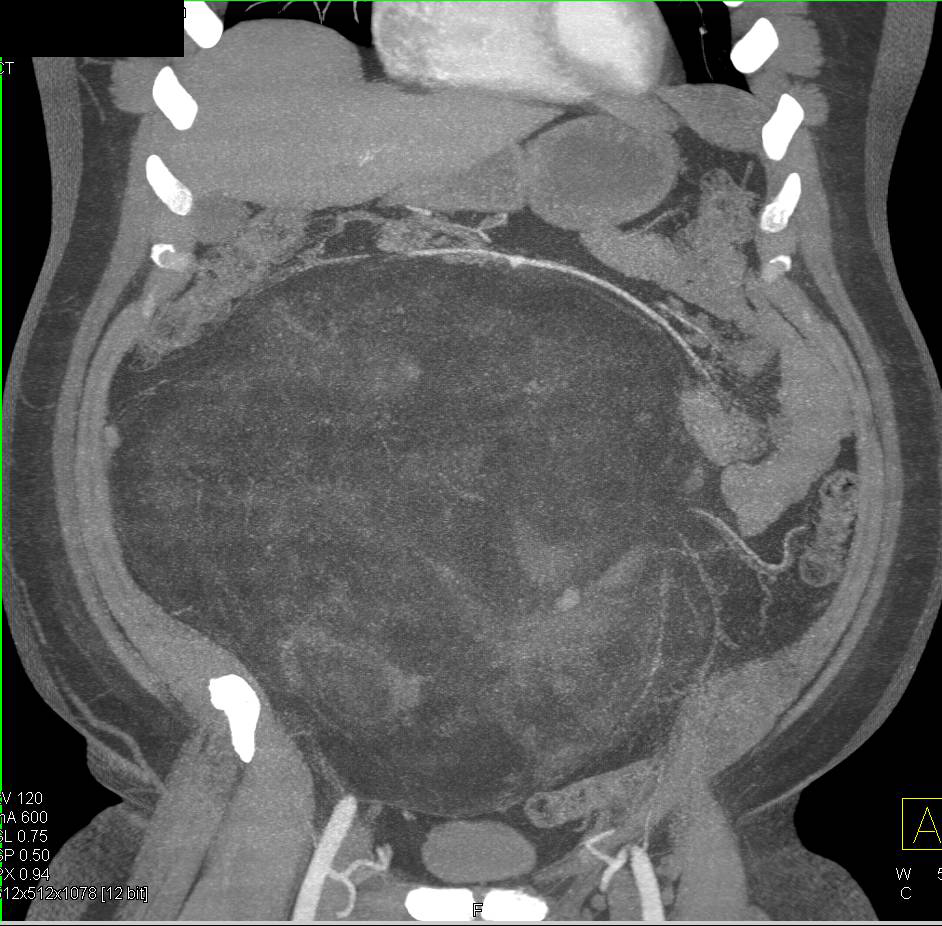
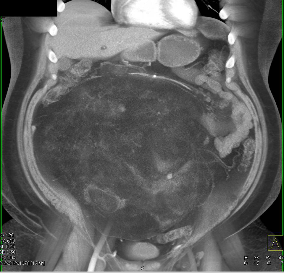
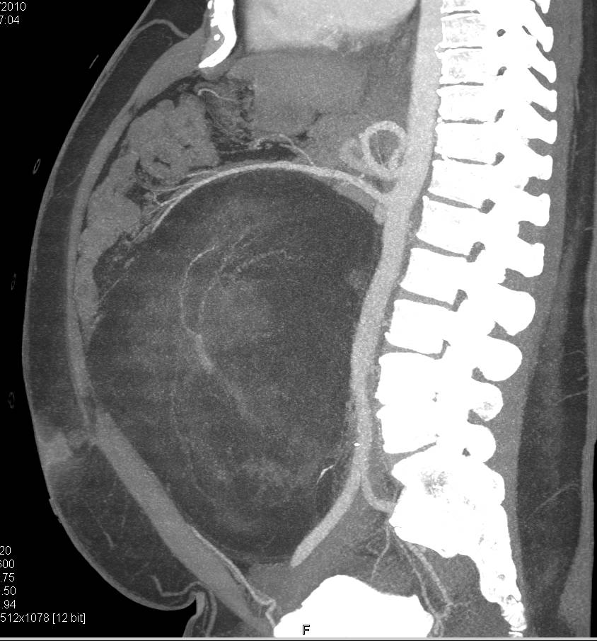
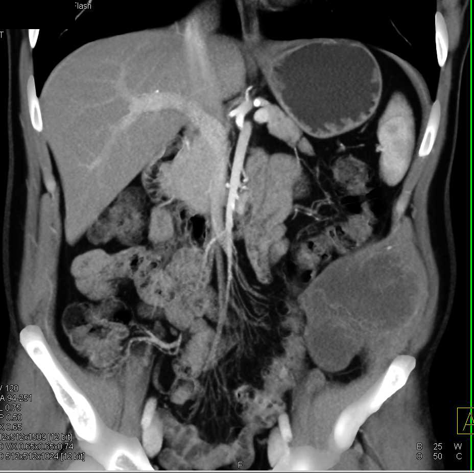
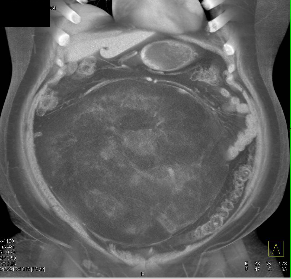
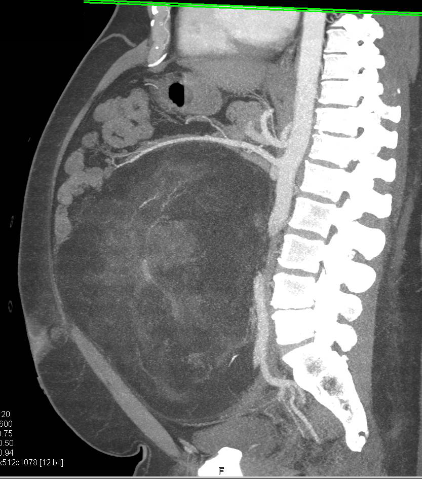
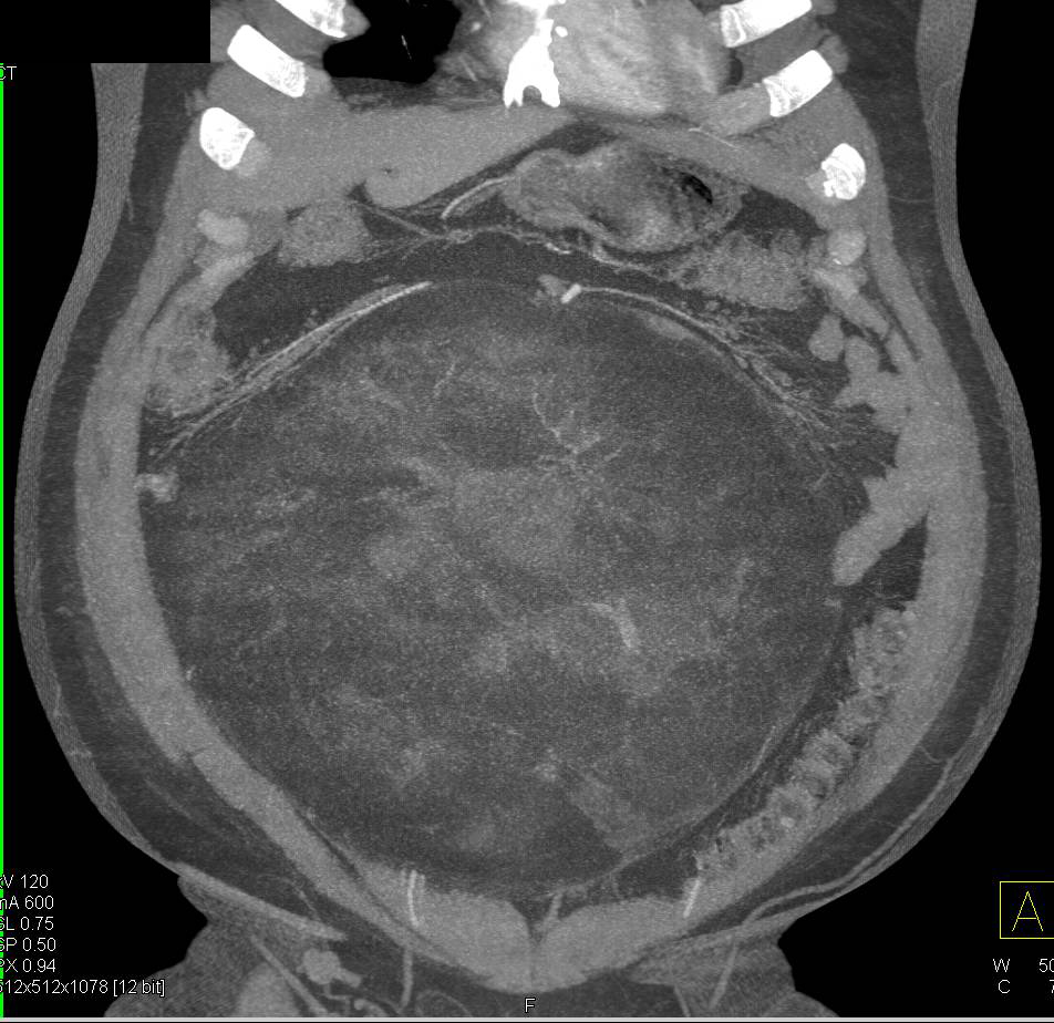
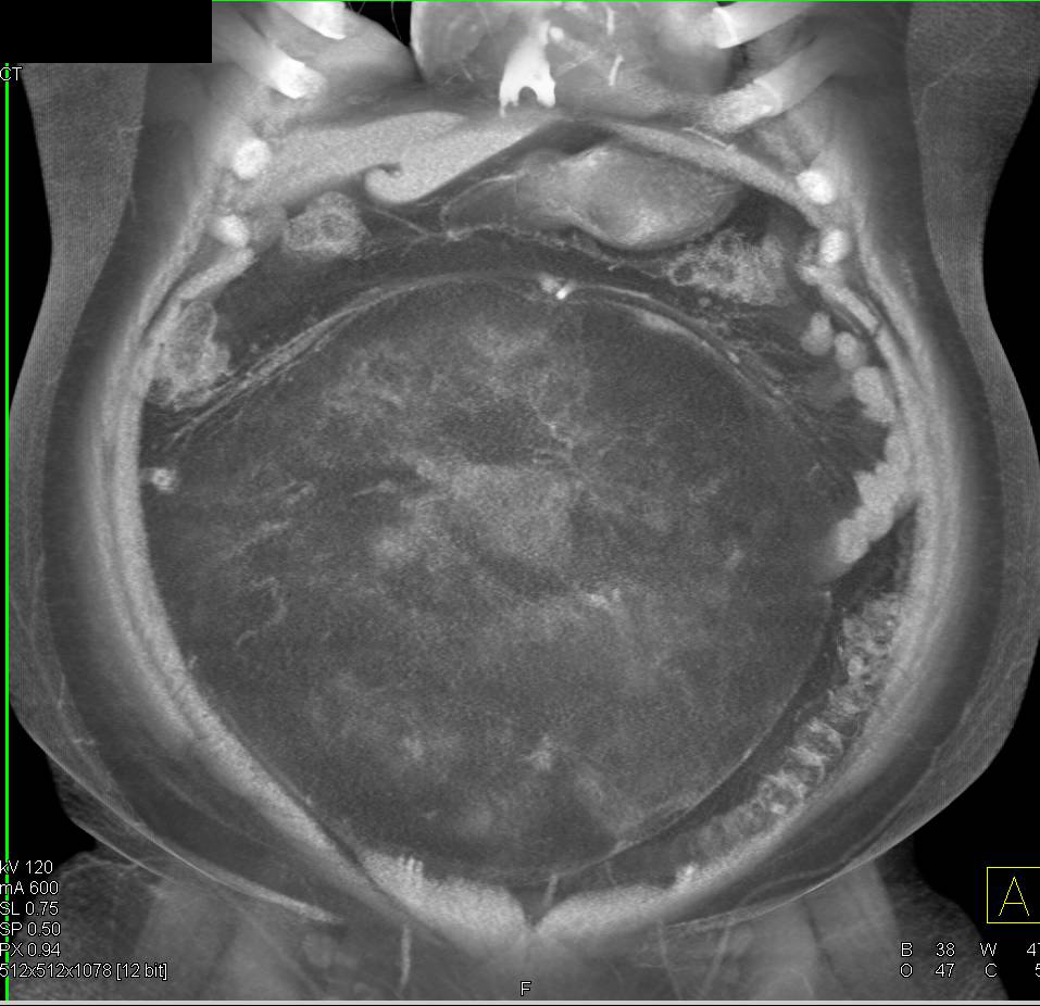
Abdominal Gastrointestinal Case Studies CTisus CT Scanning
Read This Also
- lavazza
- larioja com
- lavanguardia com
- la provincia
- ligadura de trompas
- la que se avecina
- liga nuà ez facebook
- ladera citas
- liga italiana hoy
- leiomiosarcoma
Abdominal Volume Rendering of an abdominal CT … Flickr

Source: www.flickr.com
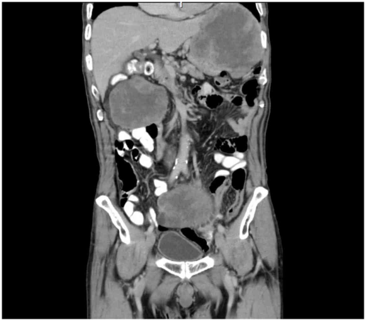
Undifferentiated abdominal wall Image

Source: radiopaedia.org
A Lowgrade Myofibroblastic in the Abdominal Cavity
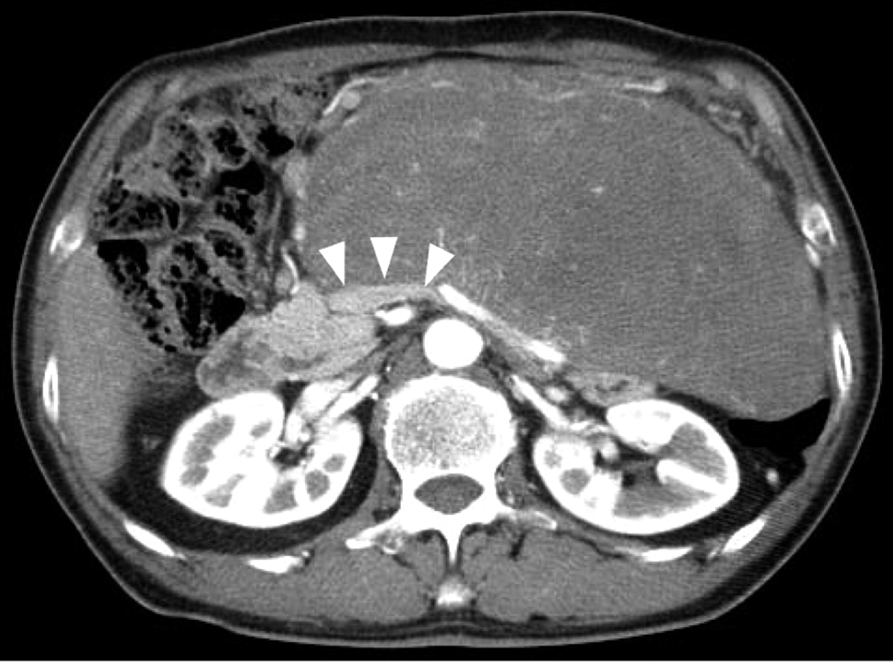
Source: ar.iiarjournals.org
What is a Retroperitoneal (with pictures)

Source: www.infobloom.com
Pictorial essay on a case of giant retroperitoneal BMJ

Source: casereports.bmj.com
result of sarcoma abdominal